Ultrasounds in Pregnancy: When and Why?

If you’re reading this, you’re most likely pregnant or looking to become pregnant. Or you could just be a reproductive health junkie like us, and curious about all things related to the female body!
No matter where you fall on this spectrum, you’ve got questions, and we’ve got answers.
Maybe not all the answers, but we can certainly clear up some confusion.
What comes to mind when you hear the word ultrasound? Funny little black and white images of what’s soon to be a full-grown baby?
Some people feel curiosity, excitement, nervousness, anxiety, and all sorts of things when they think about getting an ultrasound.
You know what they are, but do you know how they work, along with their benefits and risks?
Let’s find out!
What Exactly is an Ultrasound?
Ultrasounds, sometimes called sonograms, are used for two different purposes – therapeutic and diagnostic.
Therapeutic ultrasound uses sound waves to heal damaged tissues. You might have experienced this in a chiropractic or physical therapy office.
Ultrasounds that are done during pregnancy however are considered diagnostic.
Ultrasound technology creates images of the inside of the body using sound waves. The sound waves are at a frequency that’s above the level that humans are able to hear.
What’s An Ultrasound Like?
Ultrasounds are a quick and easy process.
They are typically performed by an ultrasound technician either in your medical provider’s office or another facility.
There are two different kinds of ultrasounds used during pregnancy.
Early on during pregnancy, you’ll most likely have what’s called a transvaginal ultrasound. This is done by inserting a wand, that is a little larger than a tampon, into your vagina. The wand is covered with a condom and lube, and doesn’t even reach your cervix.
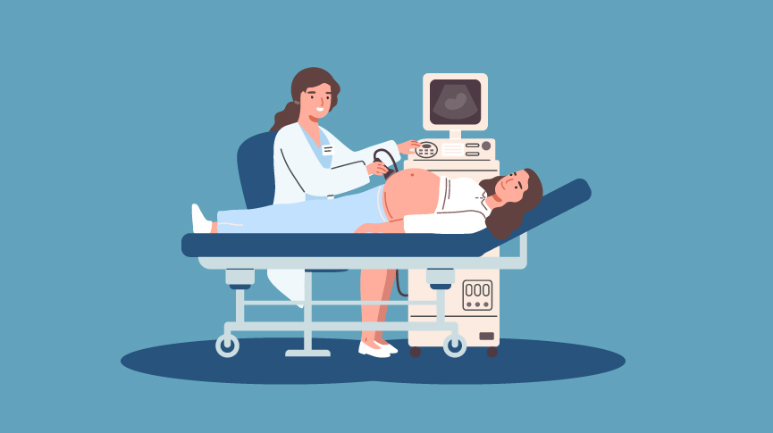
Ultrasounds that are done later on in pregnancy are typically over the skin, on the belly and pelvic area.
Before they start the imaging, they’ll apply a gel to the skin. No this isn’t to help keep your belly nice and moisturized, it’s imperative to the imaging. This gel keeps air pockets from forming between the ultrasound machine and the skin. Air pockets could block waves from going into the body, interfering with imaging.
The part of the ultrasound machine that touches your skin is called a transducer. This little doodad emits ultrasound waves and reflects them back to create two-dimensional images of tissues and organs.
The technician will pass the transducer around your belly and pelvic area to get images of your baby, and insights into its growth and development.
The whole process takes about twenty to an hour, depending on where you’re at in your pregnancy, and where your baby is positioned.
Ultrasounds in Pregnancy: When and Why
The main point of ultrasounds in pregnancy is to monitor the growth and development of your baby. That could mean a lot of different things when it comes to human development.
People typically have two ultrasounds in pregnancy. Some people may need more depending on what’s going on with them and their baby, and some people opt for none at all or just one – more on that later.
Your First Ultrasound
The first ultrasound is done in the first trimester, around week seven or eight of pregnancy. This one is often called the “dating” ultrasound, as it can help you get a clearer idea of your due date if you’re not sure.
This appointment will let you see how many babies are cooking in there – could multiples be in your future? You’ll also be able to hear your baby’s heartbeat, and see how long they are!
Genetic Screening Ultrasound
The second ultrasound is typically optional, and is done around twelve weeks gestation. This one is called the genetic screening ultrasound because it looks for any possible chromosomal abnormalities.
If your doctor does see anything during this ultrasound, they’ll most likely do further tests, and possibly refer you to a specialist.
This test is recommended for people who have a family history of genetic birth disorders. It’s important to consider whether the results of the test would impact your decision in moving forward with the pregnancy, and how it would affect your emotional health.
Halfway There Ultrasound
You’ll probably have another ultrasound at twenty weeks gestation, which is halfway through the average pregnancy.
This one is called the basic anatomy scan as your provider uses it to check if all your baby’s organs are there and are in the right place.
During this scan, you’ll be able to learn your baby’s sex – if you’d like to. Make sure to tell your technician whether or not you’d like to know.
They’ll also be able to check the amount of amniotic fluid in your uterus if your baby has any congenital heart defects, the length of your cervix, and the health of your placenta.
Keep in mind you may need further imaging, especially if you have preeclampsia, gestational diabetes, or any other factors that may impact the baby’s development.
Your standard ultrasounds are 2D images. Some people opt to get 3D or 4D ones as well. These show your baby’s features and can also give information about any spinal abnormalities and other health information.
A 4D one is similar, except that it’s a live-action shot, aka a video of your baby moving and grooving in your womb.
Are There Any Risks to Ultrasounds?
Some people believe that ultrasounds may pose a potential risk to the baby’s development because of the waves emitted.
So far, there has been little scientific evidence to back this up, and ultrasounds are considered safe for both baby and the pregnant parent.
The United States FDA (Food and Drug Administration) explains that the potential risk of ultrasounds is due to the technology’s ability to heat tissues slightly, which can produce small pockets of gas in human fluids and tissues. However, there are no known long-term effects because of this.
While we believe that it’s “Your body, your baby, your choice”, ultrasounds do offer vital information about your baby’s health and development and will be recommended by most healthcare providers.

Natasha (she/her) is a full-spectrum doula and health+wellness copywriter. Her work focuses on deconstructing the shame, stigma, and barriers people carry around birth, sex, health, and beyond, to help people navigate through their lives with more education and empowerment. You can connect with Natasha on IG @natasha.s.weiss.


It’s reassuring to know that ultrasound for pregnancy has been well-developed enough that you can already see the baby inside in either 3D or 4D that will clearly show what the baby looks likes in real-time. These endeavors in making medical services better are really commendable. I hope that when I have my own children, I could experience the joy of seeing them inside my own belly.